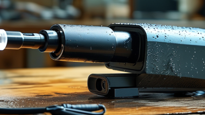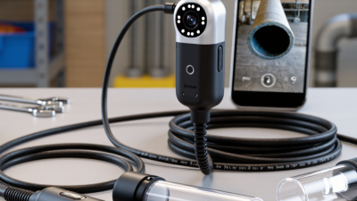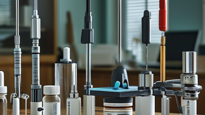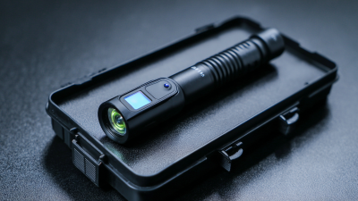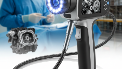

An Endo Camera is a compact imaging system used to view spaces that ordinary vision cannot reach. It combines a miniature camera, light source, lens, and flexible or rigid insertion tube. In clinical settings, doctors guide it through a natural opening or a small surgical access point. The camera sends live images to a monitor, revealing tissue color, swelling, bleeding, or unusual growths.
Professor Sir Alfred Cuschieri, a pioneer of minimally invasive surgery, described the endoscope as “the surgeon’s eye inside the body.” That image remains useful, although it is not completely perfect. An Endo Camera does not replace medical judgment. It provides visual evidence for trained professionals to interpret alongside symptoms, scans, and laboratory results.
Inside the device, light travels through optical fibers or miniature LEDs. The lens gathers reflected light, while a sensor converts it into digital signals. A processor then improves contrast and displays the image in real time. Some advanced systems offer high-definition video, angled views, zoom, or narrow-band imaging. These features can make small surface changes easier to notice.
In practice, image quality depends on more than camera resolution. Lens cleanliness, lighting, movement, and operator technique matter greatly. A small smear can hide important detail. A familiar-looking image can also mislead an inexperienced viewer. This guide explains how an Endo Camera works, where it is used, and why careful interpretation remains essential. Technology helps the clinician see more clearly, but it cannot think independently.
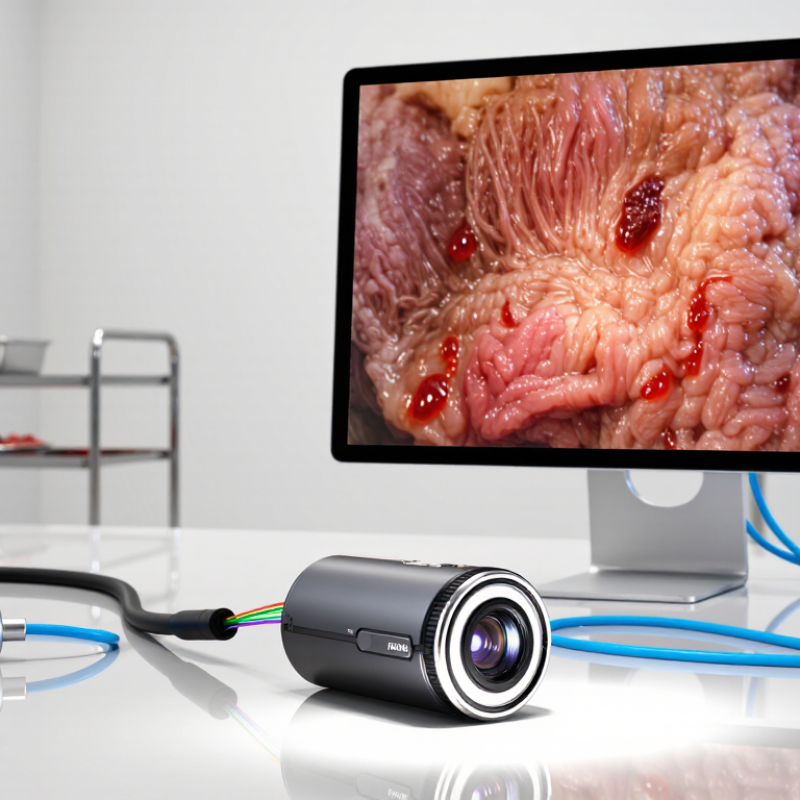
An endo camera is a miniature imaging system used inside a body cavity or narrow passage. It combines a lens, light source, image sensor, and display connection. The camera may sit at the tip of a rigid scope or inside a flexible insertion tube. In practice, the operator watches live video while steering, irrigating, or passing instruments. It is not simply a smaller video camera. Optical clarity, illumination, torque control, and sterilization affect the examination.
Rigid endoscopes use rod lenses and usually provide a bright, stable image. Flexible endoscopes use fiber optics or a distal electronic sensor, allowing navigation around curves. Capsule systems carry a camera through the digestive tract, but offer limited control and no working channel. Common outer diameters range from about 2.8 to 10 mm.
A 2.8 mm scope suits smaller passages and pediatric access. Larger 5–10 mm scopes can support stronger optics or a working channel. Diameter describes the insertion tube, not necessarily the lens or instrument channel.
A 2023 Grand View Research analysis estimated the global endoscopy devices market at roughly USD 29 billion. This growth reflects wider diagnostic use, not proof that every camera improves care.
FDA reprocessing guidance emphasizes that narrow channels and complex surfaces can retain contamination after incomplete cleaning. That warning matters in daily practice.
A practical assessment remains cautious: the thinnest scope may sacrifice brightness, stability, or instrument access. Diameter charts help, but anatomy, procedure goals, image quality, and validated reprocessing instructions must guide selection. Published market figures also vary by definition and update year. That limitation deserves attention.
An endo camera is a compact imaging system used inside narrow body passages. Its CMOS sensor converts incoming light into electrical signals. The processor then turns those signals into live video. In clinical testing, latency matters because even a slight delay can disturb hand-eye coordination. A 2024 MarketsandMarkets report identifies image quality, miniaturization, and digital integration as major endoscopy technology trends.
Light begins with high-efficiency LEDs near the camera head. They produce bright, controlled illumination without the heat of older lamp systems. The lenses guide and focus light onto the CMOS sensor. Their shape affects viewing angle, depth perception, and edge clarity. A wider lens shows more tissue, but it can reduce detail at the center. That trade-off is easy to overlook.
Tips: Check sensor resolution, LED brightness, color accuracy, and processor response together. A high-pixel sensor cannot fix poor lighting. Keep the lens clean; one thin film can create glare. The 2023 Grand View Research analysis of the endoscopy devices market also emphasizes demand for clearer imaging and less invasive equipment. Still, published specifications are not the whole story. Real performance can change with cable movement, fluid, tissue texture, and display calibration. Bench tests help, but clinical conditions remain messier.
Core Components: CMOS Sensors, LED Illumination, Lenses, and Processors
The chart shows the theoretical uncompressed RGB data rate produced by a CMOS sensor at 60 frames per second. Higher-resolution imaging creates substantially more data for the processor to handle. The lens focuses the scene onto the sensor, LED illumination provides visible light, and the processor converts, enhances, and prepares the captured signal for display or recording.
An endo camera sits at the end of a narrow, flexible or rigid scope. Tiny lenses collect reflected light from tissue, while built-in LEDs or an external light source illuminate the field. The sensor receives these photons through millions of pixels. Each pixel records light intensity and color, creating an electronic signal.
The camera then converts that signal from analog information into digital data. A processor adjusts exposure, white balance, contrast, and noise. It may also sharpen edges or separate red tones, which can help clinicians notice bleeding or unusual tissue patterns.
The processed frames travel through a cable or wireless link to a monitor. Real-time display depends on sensor speed, processor capacity, and transmission delay. A few milliseconds matter during delicate movements.
Grand View Research estimated the global endoscopy devices market at about USD 30.7 billion in 2023, with continued growth through 2030. That expansion reflects demand for clearer imaging and less invasive procedures, not merely more cameras. MarketsandMarkets has also identified image-enhanced endoscopy as a growing technology segment, although market estimates vary by definition and region. That difference deserves caution.
In practical use, image quality can fall when mucus covers the lens, light scatters, or the operator moves too quickly. I have found that brighter illumination does not always produce a better image. It can create glare and hide surface detail. Cleaning, calibration, and careful focus remain essential. The camera is advanced, but it is not independent judgment.
An endo camera is a compact imaging tool for viewing narrow, enclosed spaces. A small camera head captures images and sends them to an external monitor. Flexible or rigid shafts help the operator inspect pipes, cavities, machinery, or medical equipment. Integrated lighting is essential because these areas are often dark. Without balanced illumination, even a sharp image can look flat or misleading.
Resolution strongly affects inspection detail. A 1080p camera can show cracks, residue, and surface changes clearly during routine work. A 4K system provides finer detail, especially when the image is enlarged on a large monitor. However, more pixels do not guarantee better inspection results. Lens quality, sensor performance, lighting, and screen calibration also matter. This is easy to overlook.
Viewing angle changes what the operator can see. A 30° angle gives a more focused view, useful for examining a specific edge or opening. Wider angles, such as 70°, reveal more surrounding space and reduce repositioning. They may also create stronger distortion near the image edges. In practical use, the best angle depends on access and target size. I would not choose resolution alone. A bright 1080p image may outperform a poorly lit 4K image, which is an uncomfortable but important point.
An endo camera is a small imaging system for seeing inside narrow or hidden spaces. It usually combines a camera, light source, flexible shaft, and viewing screen. Some models also include depth control, image enhancement, and movable tips. In clinical settings, doctors may inspect body cavities, guide minimally invasive procedures, and document visible tissue changes. A sterile cover helps reduce contamination risks. The camera supports diagnosis, but it does not replace laboratory tests or professional judgment.
Industrial teams use endo cameras to inspect pipes, engines, tanks, ducts, and welds without extensive disassembly. A technician can examine corrosion, cracks, blockages, loose parts, or surface wear. The recorded images create a useful maintenance history. Time stamps, location notes, and measurements make later comparisons more reliable. Remote controls can adjust lighting, focus, or tip direction. Small details matter.
The image is not always perfect. Glare, dust, liquid, and limited angles can hide important defects. A narrow view may also make damage look smaller than it is. It needs careful handling. Skilled operators move slowly and keep the lens clean. They should verify uncertain findings with another method when safety depends on the result. This is where practice becomes important, because a clear picture can still lead to a wrong interpretation. Calibration, proper training, and honest reporting improve control and trust.