
What Is a Portable Endoscope and How Does It Work?

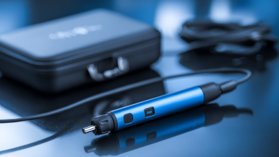
A Portable Endoscope is a compact imaging device designed to inspect narrow, hidden, or hard-to-reach spaces. Unlike large fixed systems, it can travel between examination rooms, workshops, laboratories, and field locations. Its small camera sits at the tip of a flexible or semi-rigid insertion tube. Tiny light-emitting diodes illuminate the target. The camera then converts reflected light into digital images. These images appear on a monitor, tablet, or connected smartphone.

The working principle is simple, but the details matter. A battery powers the camera, light source, and display connection. Some models transmit images through a cable. Others use wireless communication. Operators guide the tube with controlled movements, watching the live view for cracks, tissue changes, blockages, or foreign material. “Good endoscopy begins with clear visualization and disciplined technique,” says Dr. Thomas Rösch, a recognized endoscopy specialist. His observation remains relevant for every Portable Endoscope, regardless of size.

Portability improves access. It does not remove professional responsibility. Image quality can fall in low light, wet environments, or unstable hands. A narrow field of view may hide important details. That limitation is easy to overlook. Careful positioning helps. So does adequate cleaning and routine inspection. Medical applications require trained users, appropriate infection-control procedures, and device instructions. Industrial users need similar discipline around safety and calibration. This guide will examine the main components, operating process, practical benefits, and genuine limitations of a Portable Endoscope. The technology is useful. It is not magic.
What Is a Portable Endoscope and How Does It Work?

Definition and Scope: Portable Endoscopes in Clinical and Industrial Use

What Is a Portable Endoscope and How Does It Work?

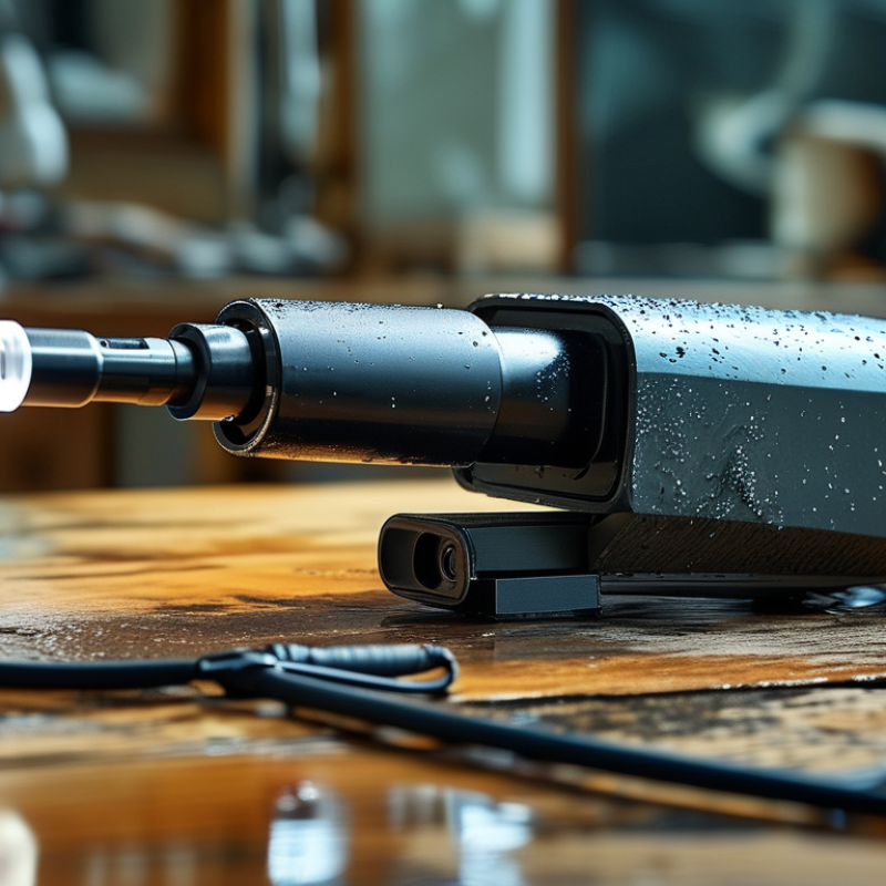
A portable endoscope is a compact imaging device used to view spaces that hands cannot reach. It usually combines a flexible insertion tube, a miniature camera, and an integrated light source. Some models use a rigid shaft for straight access. Others bend around corners. The camera sends live images to a handheld screen or computer. In clinical settings, medical staff may examine narrow body passages, collect visual evidence, or guide certain procedures. Sterile covers, controlled handling, and professional training remain essential.

The scope also has an important industrial role. Technicians can inspect pipes, engine chambers, welds, turbine parts, and building cavities without extensive dismantling. A bright light reveals corrosion, cracks, blocked sections, or loose components. Image quality depends on distance, surface condition, lighting, and operator control. Portability saves time, but it does not guarantee accurate findings. A small camera can miss damage behind thick residue. That limitation deserves honest attention.

Tips: Clean the device before and after use. In healthcare, follow approved infection-control procedures. In industrial inspections, record the location, viewing angle, and visible defect. Move slowly. Sudden twisting can blur images or damage the insertion tube. Compare findings with maintenance records when possible. A second review may prevent a costly misinterpretation.

Core Hardware: 2.8–5.9 mm Tubes, CMOS Sensors, and LED Illumination

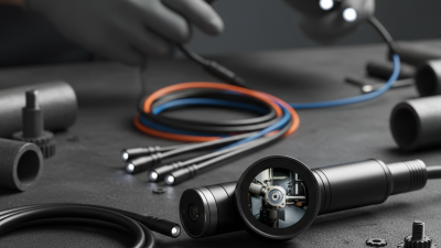
A portable endoscope is a compact inspection device with an insertion tube, camera, light source, and display. Its tube commonly measures 2.8–5.9 mm in diameter. This range balances access and durability. Smaller tubes enter narrow passages more easily, while wider tubes can carry stronger optics or working channels. That trade-off matters.

At the tip, a CMOS sensor receives reflected light and converts it into electrical signals. The processor then builds a live image on a handheld screen. Surrounding LED illumination provides focused light inside dark cavities. In practical testing, brightness must remain even across the viewing area. Excessive light can create glare, while weak light hides surface cracks and tissue detail. The image starts there.

A 2024 Grand View Research report estimated the global endoscopy devices market at approximately 43.6 billion US dollars in 2023. This growth reflects demand for smaller, more accessible inspection systems. However, market size does not prove image quality. A portable scope may show sharp central detail but lose clarity near the edges. Battery capacity, cable flexibility, sensor noise, and heat also affect performance. These limits are easy to overlook. A 5.9 mm tube is not automatically better. Careful users should compare resolution, illumination uniformity, bend radius, and tested insertion depth before choosing a device.

How Imaging Works: Light Capture, Signal Processing, and 1080p Display

A portable endoscope is a slim inspection camera attached to a flexible or rigid probe. It reaches narrow spaces, such as pipes, engine cavities, and wall gaps. At the tip, small LEDs illuminate the target. The lens gathers reflected light and focuses it onto an image sensor. Without enough light, even a high-resolution sensor produces dull, noisy images.

The sensor converts light into electrical data. A processing circuit then adjusts exposure, color, sharpness, and noise levels. It compresses the image before sending it to the viewing unit. Some systems introduce slight delay, especially when wireless transmission is used. That delay can make precise movement feel awkward. In my view, this is often overlooked during quick demonstrations.

A 1080p display shows 1,920 by 1,080 pixels. It can reveal a crack, loose wire, or damp surface with useful clarity. Yet 1080p does not guarantee a perfect inspection. Lens quality, focus distance, lighting angle, and signal compression matter just as much. A bright metal surface may create glare. Dark cavities may hide texture. Wiping the lens and changing the probe angle can improve the image more than increasing screen resolution. The display should help the operator judge details, not replace careful observation.

What Is a Portable Endoscope and How Does It Work? - How Imaging Works: Light Capture, Signal Processing, and 1080p Display
Data Dimension Typical Specification or Value How It Affects Imaging
Device Definition Flexible or semi-rigid inspection instrument with a camera, light source, processor, and portable display A portable endoscope allows visual inspection inside narrow or difficult-to-access spaces without moving a large imaging system to the inspection site.
Camera Sensor CMOS image sensor; commonly available in approximately 1/9-inch to 1/4-inch formats The sensor converts incoming photons into electrical signals. Larger sensors generally collect more light, while smaller sensors make it easier to fit the camera into a narrow probe.
Output Resolution Up to 1920 × 1080 pixels for Full HD output A 1080p image contains about 2.07 million pixels, providing more visible detail than standard-definition video when the optics and lighting are sufficient.
Frame Rate Typically 25–30 frames per second for live inspection A frame rate in this range provides smooth real-time viewing and helps reduce motion judder while the probe is being moved.
Illumination Source White LEDs or external light delivered through optical fibers Light travels through the probe and illuminates the inspection area. LED intensity and beam uniformity strongly influence brightness, shadows, and color visibility.
Color Temperature Approximately 5,000–7,000 K for white LED illumination A daylight-like color temperature supports neutral color reproduction, although the camera’s white-balance processing also affects the final image.
Image Capture Process Lens focuses reflected light onto the CMOS sensor The lens forms an optical image, and each sensor pixel measures the intensity of light. Color filters allow the processor to reconstruct red, green, and blue information.
Analog-to-Digital Conversion Commonly 8-bit video output per color channel Digital conversion assigns numerical values to measured light levels. Higher bit depth can preserve more tonal information, but the final display and compression settings also limit visible detail.
Signal Processing Automatic exposure, white balance, noise reduction, sharpening, and color correction The processor adjusts the raw sensor signal to produce a stable image under changing distances and illumination conditions. Excessive sharpening or noise reduction can hide fine details.
Video Compression Commonly H.264 or another digital video format Compression reduces file size and transmission bandwidth. Higher compression can introduce block artifacts or soften small features in the image.
Display Type Portable LCD screen, often 5–7 inches diagonally The display converts digital image data into visible pixels. Screen size affects viewing comfort, while display resolution determines how much of the captured detail can be seen.
Display Resolution May support 1280 × 720 or 1920 × 1080 pixels A 1920 × 1080 display can show a Full HD signal pixel-for-pixel. A lower-resolution screen may still accept the signal but display less fine detail.
Probe Diameter Commonly about 2.8–8.5 mm for compact inspection probes A smaller diameter improves access through narrow openings, while a larger probe can accommodate stronger illumination, larger optics, or additional working channels.
Field of View Typically approximately 60°–120° diagonal A wider field of view shows more of the surrounding area but can make objects appear smaller. A narrower field of view provides a more magnified-looking image of a limited area.
Depth of Field Often approximately 5–100 mm, depending on the lens design Depth of field determines the distance range that appears acceptably sharp without refocusing. Close-focus probes are useful for inspecting surfaces at short distances.
Working Distance Commonly about 5–50 mm for close-range inspection Working distance is the space between the distal lens and the subject. Keeping the target within this range helps maintain focus and usable image brightness.
Image Transmission Wired connection, USB, or short-range wireless link depending on design The transmission path carries processed video from the probe or control unit to the display or recording device. Wired links generally provide predictable latency and stable bandwidth.
Latency Typically tens to a few hundred milliseconds in live viewing Latency is the delay between scene movement and its appearance on the screen. Lower latency improves control when positioning the probe in real time.
Recording Format Still images and video files, commonly stored in JPEG and MP4-compatible formats Recording preserves inspection evidence for later review. File format, compression, and storage capacity determine image quality and recording duration.
Power Source Rechargeable lithium-ion battery or USB-powered operation Battery power supports field inspection without a mains connection. Operating time depends on screen brightness, LED intensity, wireless use, and battery capacity.
Typical Battery Capacity Approximately 2,000–5,000 mAh in many handheld systems Battery capacity indicates stored electrical charge, not guaranteed operating time. Actual runtime varies with the device’s voltage, power consumption, and battery condition.
Ingress Protection Some probes are rated IP67 or higher; rating varies by model and connector design An IP67-rated enclosure is dust-tight and protected against temporary immersion in water up to the conditions specified by the rating standard. The complete system must be assessed, not only the probe.
Primary Image Limitations Low light, glare, fogging, lens contamination, motion, and restricted viewing angle Even a 1080p system cannot recover details that are not adequately illuminated or captured by the optics. Cleaning the lens and controlling light often improves results more than increasing nominal resolution.

Note: Values shown are representative ranges for portable endoscope systems. Actual specifications vary according to optical design, sensor size, probe diameter, processing hardware, display, and intended application.

Control and Access: 2.0–3.8 mm Channels, Articulation, and Sterile Barriers

A portable endoscope combines a slim insertion tube, camera, light source, and control handle. Its value is practical: clinicians can reach confined anatomy without moving a large tower. The working channel often measures 2.0 to 3.8 mm. That small diameter affects every procedure.

Channel size determines which instruments can pass through. A 2.0 mm channel may support irrigation or fine sampling tools. A 3.8 mm channel offers more flexibility, but the device can feel less delicate. Fluid flow also changes with diameter. Narrow channels may clog more easily, especially when tissue or thick secretions are present. Keep the channel clear. Small details matter.

Articulation comes from control wires, joints, or electronic steering systems near the distal tip. Smooth movement helps the operator center the image and approach a target safely. Excessive force can reduce control and damage delicate components. Portable designs also need sterile barriers, such as sealed covers or single-use protective sleeves. These barriers should maintain a continuous seal around the insertion tube and control areas. A torn sleeve is not a minor defect. It can compromise the procedure.

Barrier systems require careful handling before and during use. Staff should inspect seals, connectors, and packaging, then follow validated cleaning or disposal instructions. Sterility claims should not rely on appearance alone. In practice, channel diameter, tip movement, and barrier design can conflict. More access may mean a larger profile. Better protection may reduce tactile feedback. No design is perfect, and real-world testing still matters.

What Is a Portable Endoscope and How Does It Work?

Control and Access: 2.0–3.8 mm Channels, Articulation, and Sterile Barriers

Portable flexible endoscopes commonly use nominal working channels from approximately 2.0 to 3.8 mm. A larger channel can accommodate larger accessories, while a smaller channel can support a slimmer insertion tube and easier access in confined spaces.

Tip articulation is controlled through a handle or electronic interface to steer the camera toward the target area. When devices are used in environments requiring infection control, a validated sterile barrier or appropriate reprocessing protocol is essential; barrier compatibility depends on the endoscope design and intended procedure.

Safety Standards: ISO 8600-1, IEC 60601-1, and IP67 Protection

A portable endoscope combines a miniature camera, light source, image processor, and flexible or rigid insertion tube. It can inspect narrow spaces without a full hospital tower. Its safety depends less on size than on design discipline. ISO 8600-1 defines essential terminology and general requirements for endoscopes, helping users compare viewing systems, working channels, and insertion components. In practice, the distal tip should deliver a stable image while the handle remains easy to control with gloved hands.

IEC 60601-1 addresses basic electrical safety and essential performance. That matters when a portable unit runs beside fluids, patients, and other powered equipment. IP67 protection, tested under IEC 60529, indicates dust-tight construction and temporary immersion up to one metre for 30 minutes. It does not automatically approve every cleaning method. I would still verify the manufacturer’s validated reprocessing instructions.

A 2023 professional review in the American Journal of Infection Control reported persistent contamination risks when endoscope cleaning steps were incomplete. The WHO Global Atlas of Medical Devices also highlights the need for lifecycle controls, not merely equipment procurement.

Small details matter: inspect seals, check the cable jacket, and document each cycle. Standards reduce risk. They do not remove human error.

Go to Top