What Are the Top Types of Video Otoscopes?
Choosing an ear-imaging device is not just a question of screen size. A Video Otoskop can be handheld, smartphone-connected, or designed for a fixed clinical workstation. Each type offers a different balance of portability, image control, and workflow. Some models display the ear canal on a built-in screen; others send images to a phone or computer. Small differences matter when examining a child who cannot sit still, or capturing a clear view in a dim room.
The need for reliable ear assessment is significant. The World Health Organization’s 2021 World Report on Hearing estimates that more than 1.5 billion people live with some degree of hearing loss, with that number projected to reach nearly 2.5 billion by 2050. These figures describe hearing loss, not demand for video otoscopes, but they underscore the importance of accessible ear care. A device can help clinicians view and document the eardrum. It cannot, by itself, establish a diagnosis. Image quality, lighting, safe handling, and the operator’s training all affect what can be seen. And devices differ.
This guide compares the main types of video otoscopes by design and use, including portable, connected, and clinic-based systems. It also considers practical details: whether images can be saved, how comfortably the probe is handled, and how well the device fits everyday examinations. Specifications can look impressive on a product page. Real-world usability deserves equal scrutiny.
How Video Otoscopes Are Commonly Classified
What Are the Top Types of Video Otoscopes?
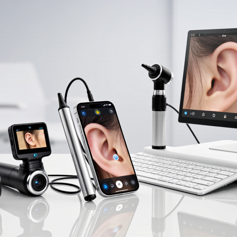
Video otoscopes are commonly classified by how they display and share images. A wired model sends live footage to a connected screen, which can make viewing steady in a clinic room. Wireless models send images to a compatible display or mobile device, reducing cable clutter but relying on battery life and connection quality. Some compact units integrate the camera, light, and screen in one handheld body.
Portability is another useful way to group them. Handheld devices suit examinations across rooms, while larger workstation systems may offer a bigger display and convenient image storage. The categories overlap. A portable scope can also be wireless, for example, so labels alone do not tell the whole story. Check image clarity, focus control, illumination, and whether the device saves still images or video. Small details matter, especially when examining a narrow ear canal.
Video otoscopes may also be described by their intended setting, such as professional clinical use or supervised teaching. They are not interchangeable with ordinary cameras: suitable lighting, cleanable contact parts, and compatible ear specula all matter. A crisp picture helps, but it does not confirm a diagnosis. Not always obvious. Device choice should fit the examination, cleaning routine, and user training; these practical limits are easy to overlook when comparing features.
Handheld Video Otoscopes with Built-In Displays
Handheld video otoscopes with built-in displays combine a viewing camera and screen in one portable instrument. Their compact design can make routine ear examinations easier to set up, especially in smaller rooms or during mobile visits. A clinician can hold the device in one hand and view the ear canal on the attached display. No separate monitor. The screen also lets a patient see what the clinician sees, which may help explain an examination in clear, concrete terms.
A built-in display is convenient, but its size and brightness matter. Check that the image remains clear under the room’s lighting and that controls are easy to reach while holding the handle. A bright exam room can cause glare. Clean the parts that contact the patient according to the device instructions, and use an appropriate single-use speculum when required. Keep the probe steady; small movements can change the view or cause discomfort. Not every ear is easy to examine. Wax, narrow canals, or patient movement may limit what appears on screen, so the image should support—not replace—clinical judgment. The screen can feel small during longer examinations, and that trade-off deserves attention when choosing a device.
Smartphone-Connected Video Otoscopes
Smartphone-connected video otoscopes pair a small camera and light with a phone or tablet, showing the ear canal on a screen. The larger view can help a clinician document visible findings or explain what they see. An LED lights the canal. A disposable speculum helps keep the camera tip from directly contacting skin, but it must be fitted correctly.
Convenience is a real advantage. A caregiver may find a live image easier to follow than instructions alone, and saved images can support a later discussion with a healthcare professional. Still, a clear picture does not guarantee a reliable assessment. Wax, movement, glare, and a narrow viewing angle can hide important details. The image may look reassuring when it is incomplete. That part is easy to overlook.
Check that the device works with the phone’s operating system and that its app displays a sharp, stable image. Clean or replace the speculum as directed, and never push the tip into the canal. Go slowly. Ear canals are delicate, and a phone screen can make people overconfident. If there is significant pain, bleeding, discharge, or sudden hearing loss, stop using the device and seek medical advice. Smartphone otoscopes can aid observation, but they do not replace a proper examination.
USB-Connected Video Otoscopes for Computer Use
USB-connected video otoscopes are designed to send a live ear-canal image to a computer. A clinician can view the image on a larger screen, adjust focus, and save stills when the device and software allow it. This can make demonstrations clearer for patients and support consistent documentation. The cable matters: it must reach the workstation without tugging on the handpiece or limiting movement.
The need for careful ear assessment is substantial. The World Health Organization’s 2021 World Report on Hearing estimates that nearly 2.5 billion people could have some degree of hearing loss by 2050, with at least 700 million needing rehabilitation services. That forecast does not measure demand for USB otoscopes, but it highlights the importance of accessible ear care. In practice, image quality depends on more than screen size. Tip fit, illumination, camera focus, and a steady hand all affect what appears on screen. Small details matter.
Computer-connected models can be useful in clinics, teaching rooms, and remote consultations where an appropriate viewing setup is available. They are not a substitute for clinical judgment. Reflections, wax, or a narrow view can hide important features. A saved image may also look different from the live view. The workflow can feel awkward at first. Check cleaning instructions, image storage settings, and patient consent procedures before routine use. WHO, World Report on Hearing, 2021.
Professional Video Otoscope Systems for Clinical Settings
Professional Video Otoscope Systems for Clinical Settings
Professional video otoscopes help clinicians inspect the ear canal and eardrum while displaying images on a screen. WHO’s 2021 World Report on Hearing projects that nearly 2.5 billion people will have some degree of hearing loss by 2050; at least 700 million may need hearing care services. This growing need makes clear, repeatable examination workflows valuable. A steady image matters. Look for adjustable lighting, dependable focus, and video capture that supports clinical documentation. A crisp picture can still mislead if wax, glare, or movement obscures the eardrum.
Tips: Check image quality on a real patient-like model before purchase. Clean or replace specula according to the device instructions and local infection-control procedures. Keep the screen positioned so clinicians can view it without awkward neck movement. Small ergonomic details matter.
In busy clinics, a durable handle, simple controls, and compatibility with existing documentation systems can save time. Confirm that images can be stored and retrieved under your clinic’s privacy and retention policies. WHO’s figures describe hearing-care needs, not demand for any specific device. That distinction matters. Video otoscopy also cannot replace clinical judgment or a complete assessment; even experienced users may need another view when anatomy or debris limits visibility.
Top Types of Video Otoscopes for Clinical Settings
Typical display and connection options vary by system. The chart shows common configurations, not universal specifications.
Handheld systems with built-in screens provide direct viewing; USB-connected systems typically display images on a computer; smartphone-connected systems use a mobile device; and cart-based systems commonly use an external monitor or computer. Actual features depend on the specific configuration.