

What Is Veterinary Endoscopy Equipment and How Does It Work?
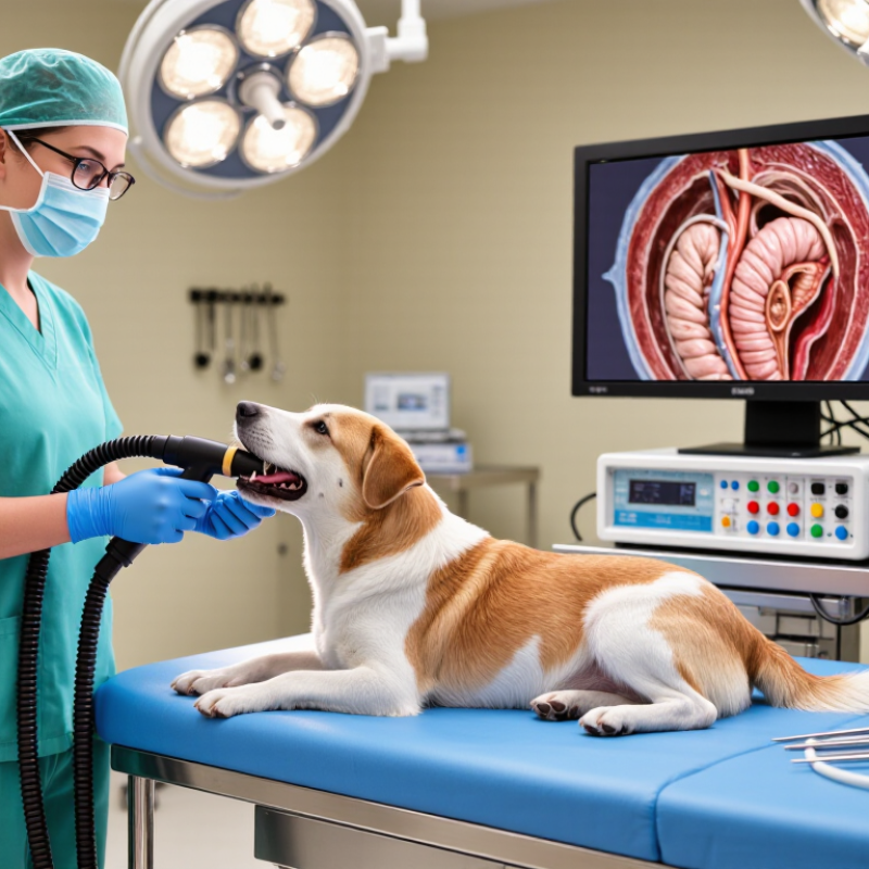
Veterinary Endoscopy Equipment helps veterinarians examine internal organs through small natural openings or carefully created access points. It usually includes a rigid or flexible endoscope, a light source, a camera, and a high-resolution monitor. Some systems also contain pumps, insufflation units, irrigation channels, and biopsy instruments. Together, these parts turn a narrow internal view into a clear image for clinical evaluation.
During a procedure, the veterinarian guides the endoscope while watching the live monitor. A flexible scope can bend through areas such as the stomach, airway, or colon. A rigid scope may provide a brighter, more stable view during joint or abdominal examinations. Working channels allow small tools to collect tissue, remove foreign material, or support other treatments. The patient commonly requires appropriate anesthesia, careful positioning, and continuous monitoring.
Precision matters.
From practical use, image quality is only one part of the process. Sterile handling, gentle insertion, and operator training strongly influence safety and diagnostic value. Equipment specifications can sound impressive, yet they may not suit every animal or procedure. That distinction deserves attention. Veterinary teams must match scope diameter, flexibility, resolution, and accessories with the patient’s size and clinical needs. Even experienced professionals may encounter limited visibility, bleeding, fogging, or unexpected anatomy. These challenges remind us that endoscopy supports clinical judgment; it does not replace it. This article explains the main components, working principles, applications, and practical limitations of Veterinary Endoscopy Equipment.
Veterinary Endoscopy Equipment: Definition, Scope, and Core Functions
Veterinary endoscopy equipment includes specialized instruments that let veterinary teams view internal structures through small openings or natural passages. A typical system combines a rigid or flexible endoscope, light source, camera, monitor, and image processor. Some setups also include irrigation, suction, insufflation, and miniature biopsy tools.
Its scope covers gastrointestinal, respiratory, urinary, nasal, and joint examinations. During a procedure, the veterinarian guides the scope while watching a magnified image on the monitor. Air or sterile fluid may gently expand a body cavity, improving visibility. Small forceps can collect tissue samples, remove foreign material, or support limited treatment. Equipment selection depends on the animal’s size, anatomy, condition, and planned procedure.
Practical reliability matters.
Veterinary staff must check image clarity, light intensity, instrument movement, and sterilization before use. An experienced operator also watches breathing, positioning, and anesthesia throughout the examination. Endoscopy can reduce tissue damage, but it is not a magic window. Blood, mucus, poor positioning, or a narrow passage can hide important findings. A clear image may still require laboratory testing and other diagnostic methods. Regular training and careful documentation help teams interpret findings responsibly, although no device removes clinical uncertainty.
| Data Dimension | Equipment or Feature | Definition | How It Works | Typical Veterinary Application |
|---|---|---|---|---|
| Definition | Veterinary endoscopy system | A medical imaging system used to inspect internal body cavities, hollow organs, and passages in animals with minimal surgical access. | A camera or optical lens at the end of an endoscope transmits magnified images to a monitor. Specialized channels can deliver instruments, irrigation, or insufflation. | Diagnostic examination, minimally invasive treatment, foreign-body removal, tissue sampling, and postoperative assessment. |
| Scope of use | Small-animal endoscopy | Endoscopic procedures designed primarily for companion animals such as dogs, cats, and similar-sized patients. | Flexible or rigid scopes are selected according to the animal’s size, anatomy, target organ, and procedure requirements. | Gastrointestinal examination, rhinoscopy, bronchoscopy, cystoscopy, otoscopy, and laparoscopic procedures. |
| Scope of use | Large-animal endoscopy | Endoscopic equipment adapted for horses, cattle, and other large veterinary patients. | Longer insertion tubes, larger working channels, and specialized access systems provide reach and instrument control in larger anatomical structures. | Equine upper-airway examination, gastrointestinal evaluation, arthroscopy, and reproductive tract procedures. |
| Visualization | Flexible video endoscope | A bendable endoscope containing a digital camera and an articulating distal tip. | Control wheels or levers steer the distal tip. Light and video signals pass through the insertion tube to a processor and display. | Gastrointestinal endoscopy, bronchoscopy, rhinoscopy, and examination of curved or difficult-to-reach passages. |
| Visualization | Rigid endoscope | A straight optical instrument containing precision lenses or a camera system. | Light is transmitted through the scope while the optical system provides a direct or camera-based view of the surgical field. | Arthroscopy, laparoscopy, thoracoscopy, otoscopy, cystoscopy, and other procedures requiring a stable viewing axis. |
| Visualization | Video processor and monitor | The electronic unit that converts the camera signal into a displayed image and may provide image recording or enhancement. | The processor receives the endoscope’s video signal, adjusts brightness and color, and sends the processed image to a medical monitor. | Real-time observation, documentation, teaching, consultation, and comparison of findings over time. |
| Illumination | Light source | A high-intensity illumination unit connected to the endoscope by a light guide or integrated optical pathway. | Light travels through fiber optics or another optical transmission system to illuminate internal tissue and anatomical spaces. | Improving visibility during inspection, biopsy, foreign-body retrieval, and minimally invasive surgery. |
| Access and control | Insertion tube and distal tip | The portion of a flexible endoscope introduced into the patient, including the steerable tip and visual components. | The operator advances, withdraws, rotates, and angulates the tube to navigate anatomy while viewing the target area on the monitor. | Targeted examination of the esophagus, stomach, intestine, airway, nasal passages, and urinary tract. |
| Working function | Instrument channel | A hollow passage within an endoscope that permits the use of accessories during a procedure. | Biopsy forceps, graspers, snares, brushes, retrieval baskets, or injection needles pass through the channel under visual guidance. | Tissue biopsy, foreign-body extraction, fluid sampling, bleeding control, and localized treatment. |
| Visualization support | Insufflation system | A system that introduces gas into a body cavity to create working space and improve visualization. | Controlled gas flow gently expands the cavity, separates tissue surfaces, and supports instrument movement during minimally invasive procedures. | Laparoscopy and other procedures in which a temporary working space is required. |
| Visualization support | Irrigation and suction | Fluid delivery and evacuation functions used to clear the lens and maintain a visible field. | Sterile fluid rinses blood, mucus, or debris from the viewing area, while suction removes excess fluid or material. | Gastrointestinal examination, arthroscopy, rhinoscopy, cystoscopy, and operative endoscopy. |
| Core function | Diagnostic imaging | Direct visualization of mucosal surfaces, lumens, cavities, and selected surgical spaces. | The endoscope provides magnified, real-time images that can reveal inflammation, ulceration, masses, strictures, bleeding, parasites, or foreign material. | Investigating vomiting, regurgitation, chronic cough, nasal discharge, urinary signs, lameness, or unexplained bleeding. |
| Core function | Biopsy and sample collection | Collection of tissue or fluid samples for laboratory and histopathological examination. | Sampling instruments are passed through the working channel and operated while the target is viewed on the monitor. | Evaluating chronic inflammation, infection, tumors, intestinal disease, airway disease, or unexplained lesions. |
| Core function | Foreign-body retrieval | Removal of ingested, inhaled, or otherwise lodged material without a large open incision when appropriate. | Graspers, snares, or retrieval baskets are guided through the endoscope to grasp and withdraw the material. | Removing selected gastrointestinal foreign bodies or material from accessible airways and body passages. |
| Core function | Minimally invasive surgery | Operative treatment performed through small access ports or natural openings using an endoscope and specialized instruments. | The scope supplies visualization while instruments perform cutting, grasping, clipping, suturing, or other controlled actions. | Laparoscopy, thoracoscopy, arthroscopy, endoscopic-assisted procedures, and selected urogenital interventions. |
| Patient safety | Anesthesia and monitoring interface | Supporting equipment used to maintain patient immobility, comfort, ventilation, and physiological stability during endoscopy. | General anesthesia or appropriate sedation is combined with monitoring of oxygenation, ventilation, circulation, temperature, and anesthetic depth. | Procedures requiring stillness, airway protection, controlled access, or extended operating time. |
| Reprocessing | Cleaning, disinfection, and sterilization workflow | A controlled process for preparing reusable endoscopes and accessories for safe subsequent use. | Immediately after use, channels and external surfaces are cleaned, rinsed, dried, and processed according to the device’s validated reprocessing requirements. | Reducing cross-contamination risk between veterinary patients and preserving equipment performance. |
| Clinical benefit | Reduced tissue trauma | A potential advantage of endoscopic access compared with selected open surgical approaches. | Natural openings or small incisions can reduce the size of the access route while maintaining direct visualization. | Potentially shorter recovery, less postoperative discomfort, and reduced disruption of surrounding tissue when clinically suitable. |
Related Posts
-
Essential Checklist for Choosing the Best Video Otoscope for Veterinary Use
-

2025 Top Veterinary Otoscope Guide for Pet Owners and Vets
-
How to Choose the Best Veterinary Otoscope for Your Practice?
-

Unlocking the Future of Medical Imaging with Hd Endoscope Camera Innovations and Insights
-
Maximizing Value with Best Veterinary Otoscope After Sales Support and Maintenance Cost Insights
-
How to Identify Quality Manufacturers for the Best Otoscope Tips You Need