
Choosing among the 10 Best Nasal Endoscope Cameras for Global Buyers requires more than comparing image resolution. A reliable Nasal Endoscope Camera must produce clear mucosal detail under uneven lighting, fit existing endoscopic systems, and support safe clinical workflows. Buyers should examine sensor quality, white balance, frame rate, lens angle, cable durability, monitor compatibility, and sterilization requirements. Small details matter. A loose connector can interrupt an examination.
Market data shows continuing demand for minimally invasive visualization. Grand View Research reported that the global endoscopy devices market was valued at approximately USD 34 billion in 2023, with steady growth expected through 2030. Fortune Business Insights also identified imaging quality, outpatient procedures, and technology upgrades as major market drivers. However, these reports use different product categories. Their figures are useful directionally, not perfectly comparable. That limitation deserves attention.
Rhinology expert Professor Heinz Stammberger helped establish modern functional endoscopic sinus surgery. His widely cited observation, “The nose is not a tube,” reminds buyers that nasal anatomy demands precise visualization and careful handling. A camera that looks impressive in a showroom may perform poorly in a narrow, wet nasal passage. Global purchasers should therefore review clinical demonstrations, ISO 13485 certification, regional regulatory status, service response times, and total ownership costs. Experience often exposes what specifications hide. The best choice is not always the most expensive model. It is the system that delivers consistent images, dependable ergonomics, and practical support across real clinical environments. Some comparisons remain imperfect, because manufacturers disclose technical data unevenly. That is exactly why independent testing and user feedback should influence the final shortlist.
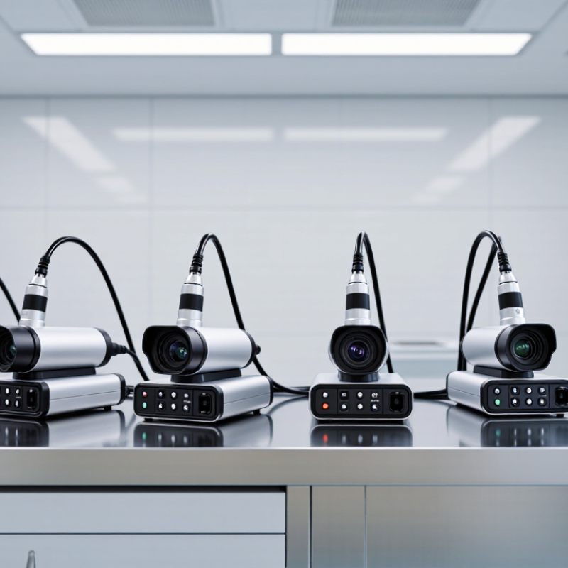
A nasal endoscope camera is a compact medical imaging system used to view the inside of the nose and nearby sinus passages.
It usually combines a slim endoscope, a tiny camera, a light source, and a monitor. The clinician guides the instrument gently through the nasal opening. Live images may show swelling, mucus, irritation, polyps, or structural differences.
Unlike a standard camera, this device must work in a narrow, moist space. Clear lighting matters. So does image stability. Some systems record photographs or video for clinical notes and follow-up comparisons.
However, a sharp image does not guarantee an accurate interpretation. Mucus, bleeding, patient movement, or an awkward viewing angle can hide important details. Professional training, careful consent, infection-control procedures, and suitable sterilization or approved single-use components remain essential. Device performance also varies, and specifications can look better on paper than during real examinations.
Tips: Check image resolution, light consistency, recording functions, and ergonomic handling. Confirm that the system fits local medical regulations and cleaning protocols. Ask whether the camera can maintain clarity after repeated clinical use. A smaller tip may improve comfort, but it can also provide a narrower field of view. That trade-off deserves practical testing, not assumptions.
A nasal endoscope camera uses a small lens, light source, and image sensor at its tip. The lens captures the nasal passage in real time. The sensor changes light into electronic signals. A processor then sends those signals to a monitor.
During an examination, a trained clinician guides the slender scope carefully through the nostril. Bright, even illumination helps reveal swelling, mucus, irritation, or unusual tissue changes. Some systems allow image capture for patient records or specialist review. However, image quality depends on more than resolution. Lens cleanliness, correct focus, stable handling, and suitable lighting matter greatly. The image is not always perfect. Moisture can blur the view, and narrow anatomy may limit movement. That practical limitation deserves honest attention.
Tips: Clean and disinfect the scope according to approved clinical instructions. Check the lens before use. Adjust light intensity gradually, rather than using maximum brightness immediately. Hold the camera steadily and avoid forcing the insertion. A short test on the monitor can reveal poor focus or image delay. Clinicians should follow local medical protocols and receive appropriate training. From an engineering perspective, comfortable grip design also matters, because hand fatigue can reduce control during longer examinations. Calibration should be checked regularly, although real-world maintenance is sometimes overlooked.
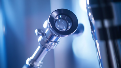
Rigid nasal endoscope systems commonly use 0°, 30°, 45°, and 70° viewing angles. A 0° scope provides a direct forward view, while angled scopes help clinicians inspect areas that are not visible along a straight line. The objective lens captures reflected light, optical elements transmit the image, and a camera sensor converts it into an electronic signal for display and recording.
10 Best Nasal Endoscope Cameras for Global Buyers?
Key Features to Compare Before Buying
Choosing a nasal endoscope camera requires more than checking pixel numbers. In practical evaluations, image clarity depends on sensor quality, lens design, lighting, and screen performance. A sharp image should reveal pale mucosa, fine vessels, and small surface changes without excessive glare. Low-latency video also matters during examination. Delayed images can make careful movement feel awkward.
Compare the camera head diameter and viewing angle with your intended procedures. A slimmer design may improve access, but it can reduce durability. Adjustable illumination helps in narrow cavities, where shadows appear easily. Check whether the system supports white balance, still images, video recording, and clear file naming. Compatibility with existing monitors or computers can reduce setup problems. Storage security deserves attention too.
Cleaning and reprocessing features should be verified before purchase. Ask whether the insertion tube tolerates the approved disinfectant and repeated cleaning cycles. Electrical safety documents, regional certifications, warranty terms, and service availability are important for global buyers. A higher resolution does not guarantee better clinical detail; I have seen bright images hide motion blur and poor focus. My own comparison process is still imperfect, especially when lighting conditions differ between test rooms. Request sample footage, inspect the connector quality, and test the controls while wearing gloves. Small details often decide whether a camera feels dependable after months of use.
Choosing among the 10 best nasal endoscope cameras for global buyers requires more than comparing image resolution. A reliable camera should provide clear views of the nasal passages, stable illumination, and natural color reproduction. Full HD may be adequate for routine examinations, while higher resolution can help document small mucosal changes. A slim distal tip, smooth handling, and responsive controls also matter during delicate procedures. The ranking should never be absolute; clinical needs differ between an outpatient room, hospital, and teaching lab.
Buyers should examine recording formats, monitor compatibility, storage security, and the availability of user instructions in their language. Adjustable white balance can improve visibility under different lighting conditions. Reusable components require validated cleaning and sterilization instructions, while disposable parts may simplify workflow but increase operating costs. Check local medical-device requirements before purchase. A camera with impressive specifications may still disappoint if replacement parts or technical support are difficult to obtain. That practical lesson is easy to overlook.
Tips: Ask for a sample clinical image, not only a catalog specification. Confirm the viewing angle, cable length, image latency, and ergonomic weight before ordering. Request documentation for cleaning, electrical safety, warranty coverage, and training. Avoid judging performance from one attractive demo. A short trial with real users can reveal glare, awkward controls, or poor image balance. Nasal endoscopy should be performed by trained professionals, with patient privacy protected during recording and storage.
Choosing a nasal endoscope camera requires more than chasing the highest resolution. Look for clear imaging, adjustable illumination, a suitable field of view, and a flexible cable. A stable monitor connection also matters during examination. For routine clinical work, recording functions should protect patient privacy and follow local data rules. The camera must suit the clinician’s hand, not only the sales sheet.
Use the device only after checking the distal tip, lens, cable, and image output. Confirm patient consent and explain each step calmly. Apply approved lubrication when appropriate, then advance gently while watching the live image. Never force resistance. Small movements reveal more than hurried insertion. WHO’s 2022 global infection prevention report estimated that 7% of hospitalized patients in high-income countries acquire at least one healthcare-associated infection; the proportion reaches 15% in low- and middle-income countries. Clean handling is not optional.
Follow the device manufacturer’s validated reprocessing instructions after every use. CDC guidance classifies instruments contacting mucous membranes as semicritical devices, requiring high-level disinfection or sterilization when suitable. Remove visible soil before disinfection, inspect channels carefully, and document each cycle. Store the camera dry, protected from dust, heat, and sharp cable bends. I have seen image quality decline from a scratched lens, not a failed sensor. That is easy to miss. Staff should also review failed examinations, because a technically correct procedure can still produce weak clinical evidence.
A practical comparison of common nasal endoscope camera configurations. Select a system according to image quality, scope compatibility, working environment, recording needs, and cleaning requirements.
| No. | Camera Profile | Recommended Use | Image Resolution | Image Sensor | Compatible Scope | Typical Working Length | Typical Field of View | Light Source | Recording / Output | Use and Maintenance Notes |
|---|---|---|---|---|---|---|---|---|---|---|
| 1 | Portable 720p USB Camera Entry Level |
Basic nasal visualization, training, and remote image sharing | 1280 × 720 pixels | CMOS | Small-diameter rigid or flexible scopes with a compatible eyepiece adapter | Approximately 175–300 mm | About 70–90° | Integrated white LED or external fiber-optic illumination | USB video; computer or compatible mobile device | Use a clean protective cover where appropriate. Do not immerse the camera head unless the manufacturer specifically permits it. |
| 2 | Portable 1080p USB Camera General Purpose |
Routine examination, documentation, and educational demonstrations | 1920 × 1080 pixels | CMOS | Rigid endoscopes and selected flexible scopes using a suitable coupler | Approximately 175–300 mm | About 70–100° | LED or fiber-optic light guide | USB, HDMI, or digital video capture | Check focus and white balance before use. Clean the optical window with approved lens tissue and a suitable optical cleaner. |
| 3 | 1080p Wireless Camera System Mobile Workflow |
Bedside viewing, teaching, and locations where cable management is important | 1920 × 1080 pixels | CMOS | Compatible rigid scopes or camera-ready flexible scopes | Approximately 175–300 mm | About 70–100° | Integrated LED or external cold-light source | Wireless display, app, or local video storage | Charge batteries before use and protect the wireless transmitter from fluids. Use an approved secure network for any patient-related transmission. |
| 4 | Full-HD Camera with HDMI Output Clinic Documentation |
Clinic examination rooms requiring a live monitor and stable video output | 1920 × 1080 pixels | 1/2.8-inch or similar CMOS | Rigid nasal endoscopes with standard camera couplers | Approximately 175–300 mm | About 70–110° | External LED or xenon-compatible light source | HDMI live output; optional USB or memory recording | Use strain relief on HDMI and power cables. Inspect the coupler, seals, and camera window before every procedure. |
| 5 | Compact 4K Camera System High Detail |
Detailed anatomy review, surgical teaching, and large-screen presentation | 3840 × 2160 pixels | 1/2.5-inch to 1/1.8-inch CMOS | Rigid scopes designed for 4K-compatible camera heads | Approximately 175–300 mm | About 70–110° | High-intensity LED or compatible external light source | 4K HDMI, SDI, or digital recording | Use a stable mount and adequate lighting. Confirm that the monitor, recorder, and cables support the selected 4K signal format. |
| 6 | 3.5–4.0 mm Flexible Video Endoscope Patient Comfort |
Flexible examination of the nasal cavity and nasopharynx | Typically 720p or 1080p | Distal CMOS sensor | Integrated flexible insertion tube; no separate rigid eyepiece required | Approximately 300–400 mm | About 90–120° | Distal LED illumination | USB, HDMI, or dedicated display unit | Follow the exact reprocessing instructions. Perform a leak test when required, and never bend the distal tip beyond its specified articulation limit. |
| 7 | 2.8–3.2 mm Pediatric Flexible System Small Diameter |
Pediatric or anatomically narrow nasal passages when clinically appropriate | Typically 720p or 1080p | Distal CMOS sensor | Integrated small-diameter flexible insertion tube | Approximately 300–400 mm | About 80–110° | Distal LED illumination | Dedicated monitor, USB, or digital recording | Use only by trained personnel. Inspect the insertion tube for damage and follow the validated cleaning and high-level disinfection process. |
| 8 | Rigid 0° Camera Configuration Straight View |
Straight-ahead inspection, anterior nasal cavity assessment, and basic procedures | 1080p or 4K, depending on the camera head | CMOS camera head | Approximately 2.7–4.0 mm rigid scope with a 0° viewing direction | Approximately 175–300 mm | About 70–90° | External fiber-optic or LED light source | HDMI, SDI, USB, or medical video recorder | Use a compatible scope coupler. Clean and disinfect the rigid scope according to its validated instructions, and protect the distal lens from scratches. |
| 9 | Rigid 30° Camera Configuration Angled View |
Viewing lateral recesses and anatomy that is not directly aligned with the nasal passage | 1080p or 4K, depending on the camera head | CMOS camera head | Approximately 2.7–4.0 mm rigid scope with a 30° viewing direction | Approximately 175–300 mm | About 70–90° | External fiber-optic or LED light source | HDMI, SDI, USB, or medical video recorder | Avoid rotating or contacting the angled distal tip against tissue. Keep the optical surface free from condensation and clean it only with approved materials. |
| 10 | Integrated Examination and Recording System Complete Workflow |
High-volume clinics needing live display, image capture, and organized records | 1080p or 4K | CMOS camera head or distal CMOS sensor | Compatible rigid and flexible nasal endoscopes | Approximately 175–400 mm | About 70–120° | Integrated LED or external medical light source | Monitor, still images, video recording, and optional network export | Create a documented cleaning workflow, restrict access to stored patient data, back up files securely, and schedule periodic inspection of cables, connectors, and light output. |