
Choosing the Best Digital Otoscope requires more than comparing camera pixels or promotional claims. Global buyers need equipment that performs reliably in real examination settings, from clinics and schools to home-care environments. A clear image, stable wireless connection, comfortable ear tips, and simple controls can matter more than impressive specifications.
This guide examines the main digital otoscope types available in 2026. These include wired USB models, Wi-Fi otoscopes, smartphone-compatible devices, professional video systems, and compact designs for mobile use. Each type offers different strengths. A wired model may provide steadier transmission, while a wireless device can improve movement around a patient. Some units include adjustable LED brightness, image capture, video recording, or interchangeable specula.
Small details matter.
A practical evaluation should also consider sensor resolution, focal distance, battery life, software compatibility, cleaning procedures, warranty coverage, and technical support. Buyers should verify whether a product meets relevant regional requirements and whether its accessories are available locally. Product descriptions can be incomplete. Even experienced purchasers may overlook app updates or connector limitations.
This overview is based on practical selection principles used in medical-device purchasing, technical comparison, and user-focused equipment assessment. It does not replace professional diagnosis or local healthcare guidance. An otoscope can help users view the ear canal, but image quality alone cannot confirm a medical condition. Careful selection remains important, yet no model is perfect for every market or user. The Best Digital Otoscope is the one that balances dependable imaging, safe handling, compliance, and long-term usability.
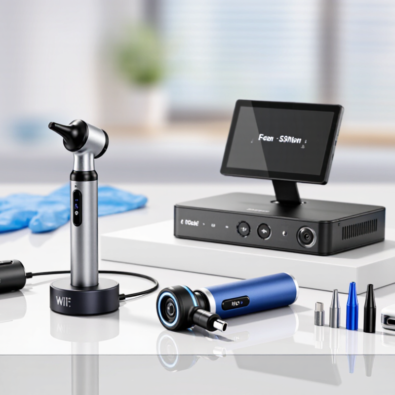
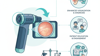
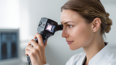
A digital otoscope is a handheld examination device with a camera, light source, and viewing screen. It illuminates the ear canal and eardrum through a small disposable or reusable speculum. The camera converts reflected light into a live digital image. Users can inspect that image on a built-in display, computer, or mobile device. Many models also save photographs or short videos for clinical records and remote consultations.
The workflow is simple, but technique matters. The operator gently positions the patient’s ear and centers the canal before capturing an image. Bright, focused lighting can reveal wax, redness, fluid, perforation, or unusual tissue changes. Image resolution, color accuracy, lens cleanliness, and stable handling affect what appears on screen. The picture can look convincing, yet it can mislead. A digital image supports assessment; it does not replace professional diagnosis.
The World Health Organization reported in its 2021 World Report on Hearing that more than 1.5 billion people live with hearing loss worldwide.
This growing need increases interest in accessible ear-screening tools. However, the same report stresses the importance of suitable services, trained personnel, and reliable referral pathways. Digital otoscopy can improve documentation and communication, especially when images accompany a patient history. It cannot measure hearing thresholds or fully assess balance disorders. Buyers should examine illumination quality, image storage security, cleaning requirements, and compatibility with existing clinical systems before selecting a device. Small details matter.
2026 Top Types Best Digital Otoscope for Global Buyers
Main Types of Digital Otoscopes for Different Examination Needs
Digital otoscopes vary by connection, image output, and examination setting. A wired USB otoscope suits clinics needing stable video, continuous power, and simple computer recording. It often works well in consultation rooms. However, cable management can slow movement around young patients.
Wireless digital otoscopes support flexible examinations in schools, home visits, and primary care. Their built-in battery should last through scheduled sessions, not just a few demonstrations. Smartphone-compatible models are convenient for quick image sharing and remote discussion, but device compatibility may require careful testing. Video otoscopes with adjustable lighting and image capture are useful for monitoring earwax, redness, swelling, or suspected fluid. High resolution helps, but it cannot replace trained clinical judgment. No device diagnoses alone.
Tips: Select the type according to examination frequency, patient age, and recording needs. Check illumination control, viewing angle, battery life, and cleaning instructions. Use disposable covers when appropriate, and disinfect reusable parts according to approved guidance. Test the software before patient use. A reliable workflow matters more than impressive specifications.
In pediatric settings, a compact tip and gentle handling are essential. For routine screening, fast startup may matter most. For specialist assessment, detailed images and secure storage deserve greater attention. I have found that buyers sometimes focus on megapixels and overlook comfort, maintenance, or staff training. That is an avoidable mistake. There is no perfect type for every facility.
| Digital Otoscope Type | Primary Examination Need | Typical Imaging Method | Common Clinical or Home-Use Features | Main Advantages | Key Limitations | Best-Fit User or Setting |
|---|---|---|---|---|---|---|
| Standard Handheld Digital Otoscope | Routine inspection of the external ear canal and tympanic membrane | Small CMOS image sensor with an integrated LED light and a viewing lens | Interchangeable disposable specula, still-image capture, video recording, and USB or wireless connection on some models | Portable, easy to share images, and suitable for general screening | Image quality depends on user positioning, ear-canal anatomy, and lens cleanliness | Primary-care clinics, school health programs, telehealth, and supervised home monitoring |
| Smartphone-Compatible Digital Otoscope | Remote consultation, patient education, and basic documentation | Camera module connected to a smartphone or tablet through a wired or wireless interface | Mobile-app preview, photo and video storage, electronic sharing, and rechargeable battery operation | Convenient for telemedicine and rapid image transmission | Compatibility, app permissions, wireless reliability, and device disinfection must be managed | Telehealth providers, community screening teams, and consumers using professional guidance |
| Video Otoscope with Large Display | Real-time examination, teaching, and examination-room collaboration | Digital camera connected to a built-in monitor or external display | Live enlarged view, freeze-frame function, video output, adjustable brightness, and image review | Multiple viewers can observe the examination at the same time | Usually less compact and may require a stable power source or dedicated workstation | ENT offices, hospitals, training centers, and procedure rooms |
| Pneumatic-Compatible Digital Otoscope | Assessment of tympanic-membrane mobility when pneumatic examination is clinically indicated | Digital camera combined with a sealed speculum system and controlled air-pressure bulb or pump | Video recording during pressure changes, improved visualization, and speculum sealing accessories | Supports visual assessment together with controlled pneumatic movement | Requires correct sealing technique and should not be used when contraindications such as suspected tympanic-membrane perforation are present | Trained clinicians, pediatric assessment, and ENT examination settings |
| Wide-Angle Digital Otoscope | Examination of larger areas of the ear canal and tympanic membrane | Wide-field optical design paired with a digital image sensor and LED illumination | Broad field of view, video capture, image documentation, and improved situational awareness during insertion | Reduces the need for repeated repositioning in suitable ear canals | Wide-angle views may provide less detail than specialized close-up imaging, depending on optical design | General examination, telehealth documentation, and clinical education |
| High-Definition Diagnostic Video Otoscope | Detailed documentation of the ear canal and tympanic membrane | Higher-resolution digital sensor with focused optics, LED illumination, and image-processing software | High-detail stills, video recording, digital zoom, adjustable illumination, and exportable patient records | Useful for comparing follow-up images and supporting specialist referrals | Higher cost, greater storage requirements, and image quality still depends on technique and focus | ENT clinics, hospitals, audiology services, and specialist teleconsultation |
| Articulating or Flexible-Tip Digital Otoscope | Improved access when the ear canal angle makes alignment difficult | Camera and illumination assembly mounted on a steerable or flexible distal section | Tip articulation, controlled insertion, live video, image capture, and interchangeable protective covers or specula | Can assist with viewing challenging canal angles under trained clinical use | More complex handling, higher maintenance needs, and increased risk of discomfort if inserted improperly | Experienced clinicians, pediatric assessment, and selected specialist examinations |
| Ear-Cleaning Digital Otoscope | Visual guidance during removal of superficial cerumen | Compact camera with LED illumination, usually positioned near a guarded cleaning accessory | Live smartphone display, removable tips, illumination control, and image or video recording | Provides visual feedback during limited, superficial cleaning | Should not replace professional cerumen management; unsuitable for deep instrumentation or suspected ear disease | Clinicians trained in ear care; consumer use only with professional instructions and safety limits |
Selection note: Digital otoscopes are intended to support visualization and documentation. Examination, pneumatic testing, foreign-body removal, and ear cleaning should be performed only by appropriately trained users, with suitable infection-control procedures and disposable or properly sterilized patient-contact components.
When comparing the best digital otoscope types, start with the examination setting. Wired USB models suit fixed consultation rooms and usually offer stable video transmission. Wireless models improve mobility but depend on battery life, signal strength, and secure data handling. Smartphone-compatible units are convenient for remote consultations, although screen quality can vary widely.
Image quality matters beyond megapixels. Check illumination uniformity, focus range, field of view, and color accuracy. A bright white LED may still create glare inside a narrow ear canal. The Lancet’s 2021 Global Burden of Disease analysis estimated that 1.57 billion people lived with hearing loss in 2019. Clear images can support earlier referral, but an otoscope cannot replace clinical judgment. That distinction matters.
Compare probe diameter, patient comfort, and cleaning requirements. A smooth, angled tip can reduce discomfort, especially for children. Disposable covers may improve hygiene, while reusable parts need documented disinfection compatibility. The World Health Organization’s World Report on Hearing projects that nearly 2.5 billion people could have hearing loss by 2050. This increases the value of practical, shareable examination tools. Still, a higher resolution does not guarantee better diagnosis. I would test the device with a dark ear model, a narrow canal, and moving video before purchase. Storage capacity, export formats, encryption, warranty terms, and compliance with relevant electrical safety standards also deserve careful review. Specifications are useful, but real handling can expose weaknesses.
How to Select the Best Digital Otoscope for Global Buyers
Selecting a digital otoscope starts with the examination setting, not the highest specification. The WHO World Report on Hearing states that over 1.5 billion people live with hearing loss worldwide. This figure may approach 2.5 billion by 2050. Buyers should therefore prioritize reliable images, simple operation, and wider clinical access.
Check image resolution under real conditions, not only laboratory lighting. A bright ear canal can expose weak automatic exposure controls. Look for adjustable LED illumination, stable focus, and a camera angle that shows the tympanic membrane clearly. A sealed, easy-to-clean surface matters in busy clinics. Disposable specula should fit securely and remain comfortable for children and adults.
Data handling deserves equal attention. Choose encrypted storage, user access controls, and export formats compatible with local medical systems. The FDA’s 2023 medical-device cybersecurity guidance supports security planning throughout a device’s lifecycle. Compliance evidence should include electrical safety testing, biocompatibility information, and regional registration documents. Ask for independent clinical validation, not only supplier claims. That takes time.
Battery life is often overstated.
Test it with continuous imaging, wireless transfer, and maximum brightness. A device promising eight hours may perform differently in cold rooms or weak networks. I would also request training materials, replacement-part availability, and response times for technical support. Buyers sometimes focus too heavily on pixel counts. That is a mistake worth reconsidering.
For global buyers, a digital otoscope is more than a camera with an LED. Its electrical design, software, patient contact parts, and intended claims affect compliance. IEC 60601-1 addresses basic electrical safety, while ISO 14971 supports medical-device risk management. In the United States, the FDA Quality Management System Regulation takes effect on February 2, 2026, aligning more closely with ISO 13485. Requirements still vary by market. A product accepted in one country may need new evidence elsewhere.
Cleaning deserves equal attention. The WHO Global report on infection prevention and control estimates healthcare-associated infections affect 7 in 100 acute-care patients in high-income countries and 15 in 100 in low- and middle-income countries. Removable patient-contact tips, clear cleaning instructions, and traceable maintenance records can reduce avoidable risk. Yet no instruction sheet is perfect. Residue can remain around the lens or hinge. Buyers should verify chemical compatibility, validated disinfection methods, ingress protection, and battery safety. The smallest gap may matter.
Tips: Request the declaration of conformity, risk-management summary, usability evidence, and local registration status. Check tip fit before patient use. Inspect the lens under bright light. Record cleaning dates and software versions. Do not rely on marketing claims alone. A practical review should include one real workflow, with gloves, limited lighting, and a busy clinic schedule. That is where weak designs become obvious.